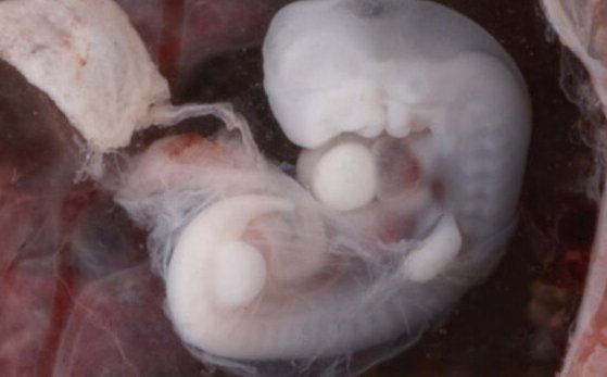
When building an embryo, timing is everything

There’s a bit of a problem in biology that’s so obvious that most biologists don’t end up thinking of it as a problem. Humans and mice (and most other mammals) all make pretty much the same collection of stuff as they develop from a fertilized egg. And they do that using a near-identical set of genes. But mice do it all in 21 days; it takes humans over 10 times longer to do it.
You might try to ascribe that to the different number of cells, but as you move across the diversity of mammals, none of that really lines up. Things get even more confusing when you try to account for things like birds and reptiles, which also use the same genes to make many of the same things. The math just doesn’t work out. How do developing organisms manage to consistently balance cell number, development time, and a static network of genes?
Biologists are just starting to figure that out, and two papers published this week mark some major progress in the field.
Getting on our nerves
One of the two sets of researchers, based in the UK, looked at the production of motorneurons, which go on to connect the spinal cords to muscles, enabling us to move. Making motorneurons takes less than a day in zebra fish, about four days in mice, and two weeks in humans—the timing differs rather dramatically. Yet the process is all controlled by an identical set of genes in these species, so it’s not an obviously genetic difference.
To figure out what was going on, they used a system in which stem cells are directed to form motorneurons. They found that, even outside the developing embryo, cells still obeyed some sort of internal clock: mouse stem cells took two to three days to form motorneurons, while human stem cells took about a week.
Why’s that happen? Maybe, the researchers reasoned, human cells don’t get as much of the signal that tells cells to develop as motorneurons. The team made some more stem cells and exposed them to a chemical that mimics that signal. This didn’t change anything. Maybe key genes in motorneuron development were regulated differently in human cells. So, they took the human version of one of these genes and put it into mouse cells. It behaved just as the mouse gene does. This indicates that the gene regulation isn’t a factor, since it just follows whatever cell it happens to be in.
So, the researchers started looking in detail at gene activity. Starting with DNA, genes get transcribed into RNAs, which are then translated into proteins. And each of these products—the RNA and the protein—have average lifetimes before they get degraded. Since the RNA and protein production seemed not to be the controlling factor, the team checked whether the RNA and proteins lived longer in human cells. They added a label to them and then shut down production, allowing the team to trace the gradual loss of the label as the protein or RNA decayed.
This showed that the RNA for key motorneuron genes were present in equivalent levels in mouse and human cells. But the protein in mouse cells lasted less than half the time that they did in human cells. While they didn’t check specific proteins, it’s possible that this could account for some of the more rapid development in mice. Another factor was cell division. When cells divide, each daughter cell gets half the proteins its parent had. They researchers found mouse cells divide faster than human cells, which will effectively reduce protein levels even further than the lower stability.
The researchers acknowledge that they haven’t checked whether any proteins specifically involved in motorneuron development are more or less stable, or whether this difference holds in other tissues or at other times. Fortunately, another research group, this one largely based in Japan, was simultaneously looking at a different tissue.
On the flank
The researchers looked at structures called somites that form along either side of the developing spinal cord. These go on to produce things like the ribs and vertebrae, along with a lot of muscles. The ribs and vertebrae are repeated structures, and somites have a similar repeated structure in the early embryo, with dozens of them forming in a typical mammal. They form in a head-to tail direction, and their formation is run like a clockwork: a set number of hours after the previous somite forms, a new one will condense out of the loose blanket of cells on the side of the developing spinal cord.
Given the topic here, it shouldn’t surprise you to hear that the clock runs at a different timing in different species: about 30 minutes in zebrafish, 90 in chickens, two to three hours in mice, and four to six hours in humans. Once again, this raises the question of why the timing of the clock can differ so much if all these species have a very similar collection of genes.
Like the other researchers, this group used stem cells from mice and humans and induced them to form somites. Again, the stem cells behaved much like the intact embryonic tissues: it took 120 minutes for mouse stem cells to start producing somite-specific genes, and 320 minutes in human stem cells.
If the cells were signaling to each other to control the timing, then it would only work if the cells were in close physical proximity. So, the authors dispersed them into a very sparse culture dish, so that few cells would have any nearby neighbors. Despite this, the onset of somite-specific gene activity retained the distinctive timing of the two different species.
As the other group did, this research team took the human version of a key somite gene and put it into mouse stem cells. This slowed down the mouse clock, but only by about 20 minutes—it was still running much faster than in human cells. When the stem cells were used to make actual mice, the clock was slow again, but the human gene was good enough to produce a healthy adult mouse.
So, the researchers started checking how the gene was used to produce a protein. And a lot of little things seemed to add up. It took about a half an hour longer for a somite-specific gene to go from first activation to producing a protein in human cells. There were also delays in the processing of RNAs (called splicing) needed before they were ready to be translated into proteins. And, as in the other study, the protein took longer to decay in human cells.
All of this would just suggest that human cells have a generally slower metabolism, which tones all of these developmental processes down. But the authors checked the stability of six other somite-specific proteins, finding that only half of them lived longer in human cells than they do in mice. There’s clearly something more complex than a general slowdown going on.
A partial answer
Really, that complexity shouldn’t be surprising. Because, while the general outlines of vertebrate development may be identical in most species, there’s a lot of significant differences—like the elaboration of a relatively large brain in humans, or an elongated tail in mice. Given these differences, it’s probably unrealistic to think that a single, tidy system would be able to handle all the timing changes needed to make these things happen.
The fact that there seems to be a lot of things feeding into the general slowdown in humans—slower cell divisions, slower protein destruction, longer RNA processing times—could allow a larger degree of flexibility. Different tissues may use different subsets of the set of potential clocks that help time their development. Unfortunately, that would mean that researchers will have to tease apart a large collection of smaller effects, and they will learn different things when they look at different tissues.
But most of biology is built upon that sort of incremental progress, which does ultimately succeed in building a big picture.
Science, 2020. DOI: 10.1126/science.aba7667, 10.1126/science.aba7668 (About DOIs).
https://arstechnica.com/?p=1707953