It’s probably not often that someone walks into your office and says, “I want you to make me a finely engineered device that makes everything worse.” Yet that seems to be a solution to creating images of large areas that still retain lots of detail.
Normally, imaging systems are a product of extremely careful engineering. I’ve heard engineers talk about polishing lenses to an accuracy of a picometer (10-12m) because that’s what you need to do to reach some image requirements. In general, the more careful you are, the better the image. But recent developments have been going the other way. It turns out that we can make use of the randomness created by rough surfaces to obtain high-quality images.
Grind that glass
So imagine a sheet of glass: you shine laser light in, and laser light comes out. It’s the most boring show on Earth. The next step is to roughen one face of the glass. After a bit of grinding (wear a mask), you have something like the translucent glass in your bathroom window. When you shine a laser light through that, what emerges is a mess of speckles and blur. We have successfully destroyed a laser beam.
The reason the laser beam is destroyed is that each part of the laser beam experiences a slightly different thickness of glass. To visualize this, let’s freeze the laser light as it passes through the glass sheet. There in front of you, you can see the electric fields of the light waves. The peaks are all lined up beautifully, and we can draw a line along that peak across the laser beam. This line is called a phase-front. Light always travels in a direction that is exactly at right angles to the phase-front.
That’s the picture before the light hits the rough surface of the frosted glass. When the light wave hits this interface, the line along the peak of the electric field is distorted and forms a replica of the surface of the glass. Now, our line becomes a jagged mess—each part of the light wave has traveled a slightly different distance through the glass. While the phase-fronts don’t fall on a nice smooth line, the light wave still travels perpendicular to the phase-front at every location. As a result, the light heads off in all directions.
Amazingly, we can repair the damage to the light wave before it even happens. The glass distorts the phase-front in the same way every time, because the surface does not change much. So, if you measure that distortion, you can insert a device that modifies the phase-front in such a way that the light wave passes through the glass undisturbed. Essentially, the light entering the glass looks blurry and horrible, and the glass corrects all the distortions that you introduced. It sends out a beautiful light beam.
This process is really cool. You can even turn your bit of frosted glass into a lens to focus light. And plenty of work has been done to use this as a method to correct for light scattering so that medical imaging using visible and near-infrared light can reach deeper into the body.
Lots of dots, all moving together
In imaging, the more detail you want to see, the smaller the imaging area you get. The limited field of view comes because you can’t get everything in focus at once. The in-focus part of an image comes from what’s called the “focal plane” of the imaging system. Unfortunately, the focal plane is not a plane—it is a curved surface, so our in-focus image comes from everything that lies on this curved surface.
The image that results, naturally, also forms a curved surface, this one on the other side of the lens. But detectors are flat, so the in-focus part of the image is the bit of curved surface that intersects (approximately) with the flat surface of the detector. When we zoom in, the surfaces become more tightly curved, so less of the surface intersects with the detector plane. Hence, you can have a large field of view, or you can see detail.
And this is where using a bit of roughened glass as a lens may beat an ordinary lens. With roughened glass, we engineer the focal plane. That, however, takes quite a bit of work and makes imaging very slow.
The trick that many researchers use is something called the memory effect. If you tilt your glass a tiny bit, all the focus points of light will move in the same direction. But tilt the glass too far and that collective motion is lost as the angles at which the reflected light comes out start to diverge. That means you can design your lens to have multiple different focuses and scan them all by rotating the glass. This creates an image that has lots of detail and a large field of view.
The problem is that the angular memory for a random surface is limited, so you need too many focal points to cover a large field of view.
The trick, it turns out, is to use glass that is kind of, but not quite, random. A group of researchers created a designer random surface. Basically, the researchers made a sheet of glass with tiny square pillars. Each pillar is the same height, but the width can take on one of three values. The result is a surface that apparently randomizes the light, but the calculations required to correct the randomization can be done ahead of time. In effect, with a relatively simple test, the researchers can quickly align the system and start using it. They estimate that their glass sheet can produce 108 focal points up to 30 degrees, compared to 1 to 2 degrees for truly random scatterers like paint or opal glasses.

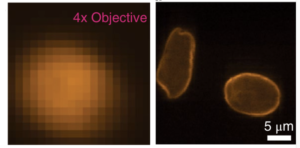
The researchers demonstrated a wide field of view and high-resolution in a spectacular way. They compared images between an ordinary microscope and their own (similar resolution and magnification). The standard microscope showed a clear image of some parasites (Giardia lamblia) with a field of view of about one millimeter. The scattering lens had the same image quality but over an area of about eight millimeters.
However, we need to recognize a limitation here. The researchers did not use multiple focal spots to image the whole field of view simultaneously. If they achieve that next step, then the true power of their microscope will be realized. There is no need for physical scanning and stitching images together to obtain an image of a large sample with high-resolution. This means that the size of the image is limited only by the dimensions of the equipment used to create the image.
Fetch forth the giant microscope
That “only” is a relative term, though. A one-meter-wide image with one micrometer resolution is probably a bit of a stretch. You will need an LCD screen with some 1019 pixels to control the light alone, never mind the rest of the setup. Nevertheless, there are plenty of cases in science and technology when reasonably high-resolution imaging over a few centimeters would be really, really useful.
Lithography—the imaging process used to print integrated circuits—springs to mind. This is a classic case of wanting both a large field of view and a high-resolution. A scattering lens is not going to replace the highest-end lithography systems. But plenty of chips are printed at much coarser resolution, and they may (eventually) benefit from this technology.
At the moment, though, this is a lab setup with all the foibles associated with its “built by a graduate student” status. In other words, it works brilliantly but is probably more temperamental than a confused and angry bull. However, if engineers from any of the bigger microscope manufacturers notice this development (which they probably will), you should start seeing scattering lenses turn up in commercial microscopes in a couple of years.
Nature Photonics, 2018, DOI: 10.1038/s41566-017-0078-z (About DOIs).
https://arstechnica.com/?p=1251921

