One of the best-known poems by Gerard Manley Hopkins opens with a tribute to the phenomenon of iridescence. It’s represented by the colorful wings of kingfishers and dragonflies in Hopkins’ poem, but iridescence can also be found in the wings of cicadas and butterflies, in certain species of beetle, and in the brightly colored feathers of male peacocks. Now, a team of researchers at MIT have captured on video the unique structural growth of butterfly wings—continuously, as a butterfly develops inside its chrysalis—for the very first time. The researchers described their findings in a new paper published in the journal Proceedings of the National Academy of Sciences.
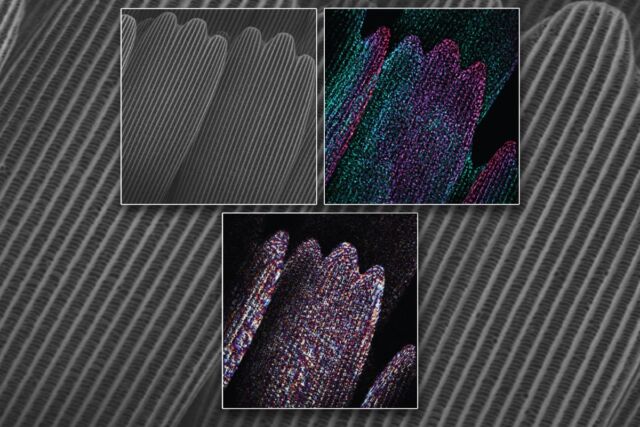
As I’ve written previously, the bright iridescent colors in butterfly wings don’t come from any pigment molecules but from how the wings are structured. It’s a naturally occurring example of what physicists call photonic crystals. The scales of chitin (a polysaccharide common to insects) are arranged like roof tiles. Essentially, they form a diffraction grating, except photonic crystals only produce certain colors, or wavelengths, of light, while a diffraction grating will produce the entire spectrum, much like a prism.
Also known as photonic bandgap materials, photonic crystals are “tunable,” which means they are precisely ordered in such a way as to block certain wavelengths of light while letting others through. Alter the structure by changing the size of the tiles, and the crystals become sensitive to a different wavelength. (In fact, the rainbow weevil can control both the size of its scales and how much chitin is used to fine-tune those colors as needed.)
Even better (from an applications standpoint), the perception of color doesn’t depend on the viewing angle. And the scales are not just for aesthetics; they help shield the insect from the elements. There are several types of manmade photonics crystals, but gaining a better and more detailed understanding of how these structures grow in nature could help scientists design new materials with similar qualities, such as iridescent windows, self-cleaning surfaces for cars and buildings, or even waterproof textiles. Paper currency could incorporate encrypted iridescent patterns to foil counterfeiters.
Butterfly wings have long fascinated scientists, ever since the first documentation of the growth of said wings back in 1938. We now have much more advanced imaging techniques, shedding further light on this complex process. “Previous studies provide compelling snapshots at select stages of development; unfortunately, they don’t reveal the continuous timeline and sequence of what happens as scale structures grow,” said co-author Mathias Kolle, a mechanical engineer at MIT. “We needed to see more to start understanding it better.”

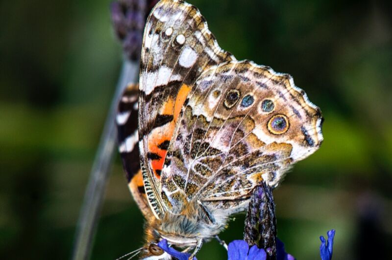
The team raised batches of painted lady butterflies (Vanessa cardui) in the laboratory, carefully monitoring the larvae housed in individual containers until the larvae molted their skins. Once the caterpillars were encased in a chrysalis and the final metamorphosis into butterflies had begun, the researchers set about recording the process. They relied upon a couple of surgical approaches to get an inside view of the wing development in the pupae.
First, the researchers exposed the forewing by removing part of the cuticle with a scalpel; the pupae were anesthetized for this process. Then they placed a thin glass coverslip over the excised area with a bioadhesive and sealed it with a handheld dental light-curing lamp.
To image the hindwings, the MIT team grasped both the chrysalis cuticles and the forewing and folded them in the direction of the head. The hindwing and forewing were separated with a strip of dental composite. Once again, the researchers used a glass coverslip to protect the exposed wing and provide a window into the chrysalis, sealing the window in place with dental composite.

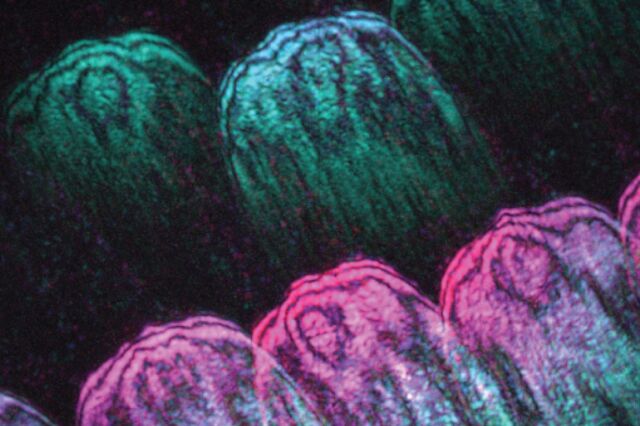
However, the researchers needed a special kind of imaging to capture the wing formation, since simply shining a wide beam of light on the wing could damage the cells. The solution: speckle-correlation reflection phase microscopy, which involves shining many tiny points of light on specific points on the wing.
“A speckled field is like thousands of fireflies that generate a field of illumination points,” said co-author Peter So, one of three experts in this imaging type of imaging who collaborated on the experiments. “Using this method, we can isolate the light coming from different layers, and can reconstruct the information to map efficiently a structure in 3D.”
https://arstechnica.com/?p=1814777

