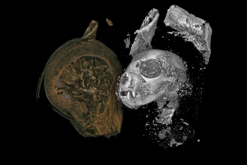
The ancient Egyptians mummified animals as well as humans, most commonly as votive offerings to the gods available for purchase by visitors to temples. Many of those mummified remains have survived but are in such a fragile state that researchers are loath to disturb the remains to learn more about them. Now an inter-disciplinary team of scientists has managed to digitally “unwrap” three specimens—a mummified cat, bird, and snake—using a high-resolution 3D X-ray imaging technique, essentially enabling them to conduct a virtual postmortem, according to a new paper published in the journal Scientific Reports.
Studying fragile ancient artifacts with cutting-edge imaging technology confers a powerful advantage on archaeological analysis. For instance, in 2016, an international team of scientists developed a method for “virtually unrolling” a badly damaged ancient scroll found on the western shore of the Dead Sea, revealing the first few verses from the book of Leviticus. The so-called En Gedi scroll was recovered from the ark of an ancient synagogue destroyed by fire around 600 CE.
In 2019, we reported that German scientists used a combination of cutting-edge physics techniques to virtually “unfold” an ancient Egyptian papyrus, part of an extensive collection housed in the Berlin Egyptian Museum. Their analysis revealed that a seemingly blank patch on the papyrus actually contained characters written in what had become “invisible ink” after centuries of exposure to light. And earlier this year, we reported that scientists had used multispectral imaging on four supposedly blank Dead Sea Scrolls and found the scrolls contained hidden text, most likely a passage from the book of Ezekiel.
Now scientists are applying advanced imaging methods to the study of mummified remains. Early techniques, dating as far back as the 1800s, were quite intrusive, usually involving the unwrapping of the mummy to study the bones and any artifacts wrapped up with the remains. They could often yield insight into wrapping techniques and the mummification process (thanks to chemical analysis), but they also resulted in damage to, or destruction of, the remains. These days, non-invasive techniques are heavily favored, such as polarized light microscopy, conventional radiography, and medical X-ray computed tomography (CT).
However, while the latter is an improvement over 2D radiography in terms of capturing volumetric (3D) qualities, medical CT also has lower resolution. Micro CT brings high-resolution capability to 3D images. It involves combining several radiographs to build a “tomogram” (3D volume image), which can then be 3D printed or analyzed in a virtual reality setting. The technique is commonly used by scientists for imaging the internal structure of materials at the microscale. Previously, Micro CT had been used to image a mummified falcon, enabling researchers to determine its likely last meal and to image a mummified severed human hand.
“Using micro CT we can effectively carry out a postmortem on these animals, more than 2,000 years after they died in ancient Egypt,” said co-author Richard Johnston of Swansea University. “With a resolution up to 100 times higher than a medical CT scan, we were able to piece together new evidence of how they lived and died, revealing the conditions they were kept in, and possible causes of death. Our work shows how the hi-tech tools of today can shed new light on the distant past.”
-
Mummified cat.University of Swansea
-
Mummified bird of prey.University of Swansea
-
Mummified snake.University of Swansea
-
Micro CT visualization of the mummified cat head.University of Swansea
-
Digitally dissected lower jaw (mandible) and teeth of the mummified kitten. Reveals fractures and unerupted mandibular first molars (red) indicating it was a kitten at the time of death.University of Swansea
-
Skeletal remains and measurements of a mummified Eurasian Kestrel.University of Swansea
-
The coiled remains of an Egyptian Cobra, undisturbed for thousands of years. Digitally dissected and revealed beneath the wrappings.University of Swansea
-
The skull of a mummified Egyptian Cobra, with mouth open, revealed by X-ray microtomography.University of Swansea
-
Micro CT visualization of the mummified snake.University of Swansea
Swansea University maintains a collection of mummified specimens, and the team selected three animal remains that varied in both size and shape: a cat, a bird, and a snake. The cat head was decorated with a painted burial mask and was wrapped separately from the body, likely after mummification. The bird of prey mummy was intact except for a severed leg protruding from the bottom of the wrapping. The mummified remains of the snake were not definitely identified as a snake until a 2009 radiograph, courtesy of a local veterinary clinic, revealed it to be coiled up inside.
Using micro CT, the team was able to image the remains in extraordinary detail, including small bones and teeth, desiccated soft tissues, and mummification materials. For instance, the researchers were able to determine that the mummified cat (likely the domestic Felis catus species) was actually a kitten, roughly five months old, after identifying teeth within the jaw bone that hadn’t yet emerged. Examination of the cat’s body showed an unfused distal epiphysis, further evidence of its young age. They even determined a likely cause of death: the separation of the vertebrae was consistent with strangulation.
The bird likely belonged to the Eurasian kestrel family, based on virtual bone measurements. As for the snake, it was identified as a mummified young Egyptian Cobra. There was evidence of kidney damage (calcification in particular) and gout, suggesting that the animal had not been kept in very good conditions and likely didn’t get enough water. There were multiple bone fractures, consistent with the snake being killed by a strong whipping motion while being held by the tail.
There was also evidence of the mouth opening being filled with resin (most likely natron) to render the snake harmless. Alternatively, the authors suggest that the material may have been placed in the mouth as part of an “opening of the mouth” mummification procedure. “The latter is supported by the fact that the snake’s jaw is wide open, an unlikely final position without some intervention to prize open and maintain separation of the upper and lower jaws,” the authors wrote. “There is also clear trauma to the jaw bones and teeth, which has been observed in human mummies that have undergone the opening of the mouth procedure.”
If so, this would be the first evidence for this practice being applied to a snake, although historical texts suggest a similar practice for the Apis bull, involving placing myrrh and natron under the tongue to slow down decomposition. In short, said co-author Carolyn Graves-Brown from the Egypt Centre at Swansea University, “Our findings have uncovered new insights into animal mummification, religion, and human-animal relationships in ancient Egypt.”
DOI: Scientific Reports, 2020. 10.1038/s41598-020-69726-0 (About DOIs).
https://arstechnica.com/?p=1700405

