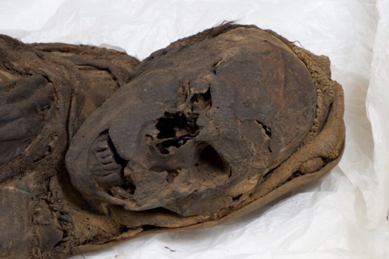
An international team of scientists used CT scanning to conduct “virtual autopsies” of three South American mummies and found evidence of fatal trauma in two of them, according to a recent paper published in the journal Frontiers in Medicine. One of the mummies had clearly been hit on the head and stabbed, possibly by two assailants, while the other showed signs of massive cervical spine trauma. The third female mummy also showed signs of trauma, but the damage was inflicted post-mortem. The study is part of ongoing efforts to determine the frequency of violence in prehistoric human societies.
According to the authors, there is a large database of ancient Egyptian mummies and skeletons that show signs of having suffered a traumatic injury, but there is far less data for South American mummies, many of which formed naturally and are exceptionally well-preserved. Nonetheless, evidence of fatal trauma has been reported previously in a few cases, such as a pre-Columbian skull from the Nasca region showing rotational trauma to the cervical spine and accompanying soft tissue bleeding into the skull. An almost complete female mummy showed signs of facial bone fractures consistent with massive strikes from a weapon, as did the skull of a mummified male infant.
An extensive 1993 survey used conventional X-rays to analyze 63 mummies and mummy fragments, 11 of which showed signs of trauma to the skull. But those mummies came from different locations, populations, and time periods, making it difficult to draw general conclusions from the findings. Last year, researchers looked for signs of violence in the remains of 194 adults buried between 2,800 and 1,400 years ago in the Atacama Desert of northern Chile, 40 of which appeared to have been the victims of brutal violence.
The authors of this most recent paper have combined expertise in anthropology, forensic medicine, and pathology and relied upon CT scanning technology to reconstruct the three mummies under investigation. “The availability of modern CT scans with the opportunity for 3D reconstructions offers unique insight into bodies that would otherwise not have been detected,” said co-author Andreas Nerlich, a pathologist at Munich Clinic Bogenhausen in Germany. “Previous studies would have destroyed the mummy, while X-rays or older CT scans without three-dimensional reconstruction functions could not have detected the diagnostic key features we found.”
-
Front and side view of the “Marburg Mummy”A-M Begerock et al., 2022
-
CT scan of the Marburg mummy’s thorax showing evidence of tuberculosis.A-M Begerock et al., 2022
-
The right zygomatic fracture of the Marburg mummy.A-M Begerock et al., 2022
-
CT cross section of Marburg mummy’s thorax showing stabbing wound reaching toward the aorta.A-M Begerock et al., 2022
The first specimen Nerlich and his colleagues analyzed is known as the “Marburg Mummy,” a mummified male housed at the Museum Anatomicum of the Phillips University in Marburg, Germany. (Acquisition records describe it as a “female mummy,” so someone at the time missed the mummy’s male genitalia.) The man was likely between 20 and 25 when he died and stood roughly 5 feet, 6.5 inches (1.72 meters) tall. He was buried in a squatting position, and given the nature of the goods buried with him, he likely belonged to a fishing community of the Arica culture in what is now northern Chile. There was prior scarring of the lungs, indicating the man suffered from tuberculosis, and he had well-preserved but crooked teeth. Radiocarbon dating indicates he died between 996 and 1147 CE.
https://arstechnica.com/?p=1880352

