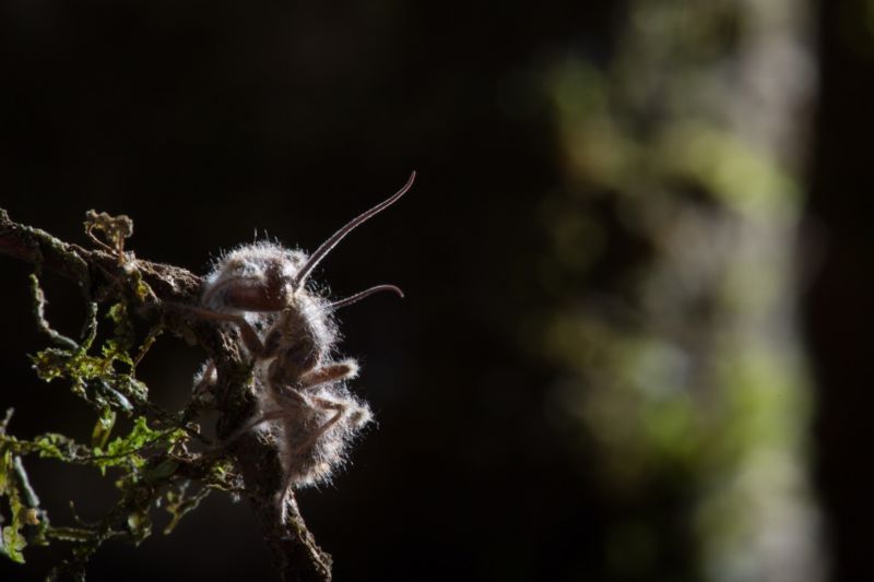
Pity the poor unsuspecting carpenter ant who unwittingly becomes infected with spores scattered by a parasitic fungus in the Cordyceps genus. The spores attach to the ant and germinate, spreading through the host’s body via long tendrils called mycelia. Cordyceps essentially turns its host into a zombie slave, compelling the ant to climb to the top of the nearest plant and clamp its tiny jaws in a death grip around a leaf or twig.
The fungus then slowly devours the ant, sprouting through its head in one final indignity. Then the bulbous growths on the ends of the mycelia burst, releasing even more spores into the air, to infect even more unsuspecting ants. It’s not a great way to go: the entire process can take four to 14 days.
There are more than 400 different species of Cordyceps fungi, each targeting a particular species of insect, whether it be ants, dragonflies, cockroaches, aphids, or beetles. The zombification aspect has made the fungus a favorite of nature documentaries. It has also worked its way into popular culture, such as the zombie-apocalypse video game, The Last of Us (2013), in which a parasitic fungus mutates so that it also infects humans. But scientists are keen to study Cordyceps to learn more about the origins and intricate mechanisms behind these kinds of pathogen-based diseases.
Parasitic puppetmaster
David Hughes, an entomologist at Penn State University, has been studying the fascinating relationship between carpenter ants and their parasitic partner, Ophiocordyceps unilateralis, for years, in hopes of learning more about how the fungus controls its doomed host. Prior research showed that the zombification might be due to the release of a special chemical that causes the muscles in the infected ants’ mandibles to contract forcefully for that death-grip bite.
Back in 2017, Prof. Hughes and his team scanned ultra-thin slices of infected ants under a powerful microscope to build a 3D model, painstakingly marking which parts were ant and which were fungus. That gave them a much more detailed look at what was happening structurally at the cellular level. They found a surprisingly high percentage of fungal cells in the ants’ bodies. The cells were concentrated directly outside the brain without ever penetrating the brain.
Instead, the fungal cells formed an elaborate, interconnected 3D network, enabling them to communicate with each other and exchange nutrients. They essentially cut the brain off from the rest of the ant’s body, so the networked cells can control its behavior. As Ed Yong wrote in The Atlantic, “The ant ends its life as a prisoner in its own body. Its brain is still in the driver’s seat, but the fungus has the wheel.”
“Its brain is still in the driver’s seat, but the fungus has the wheel.”
Now, the Hughes lab is back with a new paper in Experimental Biology. This latest study “gives us a broader picture of the fungus-host interactions that are occurring in the mandible [jaw] muscles of infected ants at the time of biting,” said Penn State’s Colleen Mangold, a co-author on the paper. Those interactions are likely what’s behind that death-grip bite, since the fungus doesn’t actually directly attach to an infected ant’s brain. Rather, it breaks apart the membrane that covers jaw-muscle fibers, causing contractions strong enough to damage or destroy the muscle filaments that slide past each other when the muscles contract.
Since this particular fungus thrives in hot, humid environments (Brazil or South Carolina), the U-Penn scientists recreated a similar environment in the lab. They collected spores from infected ants and injected them into healthy ants in the lab. The trick here was determining the correct spore dosage.
“If we inject with too few spores, the ant can fight off the infection,” said Mangold. “However, if we inject too many, the ant could die shortly after inoculation with no behavioral change. So we had to find a dosage somewhere in between.”
The lab environment was designed to resemble free-roaming conditions, not an ant colony nest. “The fungus is unable to develop to a mature, infectious stage inside ant nests,” said Mangold. “This is probably due to the fact that healthy ants sometimes physically remove infected ants from the nest, and/or conditions within the nest—like humidity or temperature—are not optimal for fungal growth. When an infected ant is away from the nest, the fungus can grow and mature, and infectious spores can be released.”
Muscles under the microscope
-
SEM image of (A) uninfected and (B, C, D) infected mandibular muscle in a carpenter ant.C.A. Mangold et al.
-
Motor neurons and neuromuscular junctions are maintained in infected mandibular muscle at the time of biting.C.A. Mangold et al.
-
SEM images of vesicle-like particles (indicated with arrows) on fungal cells.C.A. Mangold et al.
As the infected insects were dying, Mangold et al. froze them and removed the jaw muscles for preservation. Then they studied those tissues under a scanning electron microscope (SEM). Those images clearly showed the fungus filaments penetrating the muscle tissue, but the neuromuscular junctions—where nerve signals from the brain would enter the muscles to control their movement—remained intact, indicating that the fungus is not directly affecting an infected ant’s brain. The team also observed strange vesicle-like particles attached to infected tissue, although it’s not clear whether those are being produced by the fungus or the host ant.
The team will next attempt to isolate the bead-like vesicles to learn a bit more about them. “We want to know if they come from the fungus or the host and what is packaged inside them,” said Mangold. If the vesicles come from the fungus, that would suggest whatever is inside plays a role in the muscle contraction—perhaps by secreting some substance that causes spasms in the muscle—or mediates the communication between fungal cells.
If the vesicles come from the host ants, the contents could be an immune response of some kind. Either way, “Understanding more about these vesicles may help provide insight into the host-pathogen interactions that may contribute to the death-grip,” said Mangold.
Carpenter ants aren’t the only species who are targets for this kind of zombie-like infection; they’re just one of the most well-known. “There are a number of other systems where we see behavioral changes in hosts infected by a microbe,” said Mangold, pointing to ants infected with Pandora formicae as one example. “However, the specific mechanisms underlying host behavioral change may differ between the two systems. That’s why it will be interesting to study each system and see where our findings are similar and where they are different.”
DOI: Journal of Experimental Biology, 2019. 10.1242/jeb.200683 (About DOIs).
https://arstechnica.com/?p=1537389

